Myth: Dental X-rays expose you to a lot of radiation
Fact: Dental X-rays use a small, controlled dose, and digital sensors require a lower dose than traditional film.
General Dentistry · Merrick, NY
Many dental problems start where no one can see them: between the teeth, under old fillings or deep in the bone around the roots. At Modern Dental Hub in Merrick, NY, we use digital X-rays and 3D imaging (CBCT) to see those areas clearly, so that our dentists can diagnose accurately and plan treatment with precision.
Digital X-rays are part of routine care for almost every patient, but they are especially valuable if you:
X-rays are taken only when they add information the dentist needs. If you are pregnant or think you may be, tell us; we postpone routine X-rays and take only what cannot wait.
A dental X-ray is an image created when a small, controlled dose of radiation passes through the teeth and jaw. Digital X-rays capture that image on an electronic sensor instead of film and send it straight to a screen.
At Modern Dental Hub we use two kinds of digital imaging:

Digital images appear on the screen within seconds. Your dentist can enlarge them and point out exactly what they see while you watch. Digital sensors need a lower radiation dose than traditional film. Images are stored in your chart, so they can be compared visit to visit and shared with a specialist if you are referred.
For fillings, X-rays confirm how deep a cavity is and whether a filling or a crown is the right choice. For dental implants, a 3D scan measures the height, width and density of the bone before anything is planned. For root canal treatment, images show the shape and number of root canals and whether infection has reached the bone. For extractions, they show root shape and nearby structures so that the tooth can be removed safely.
Your dentist decides which images are needed. We take X-rays based on your history, your symptoms and the treatment being planned.
Protection. You wear a lead apron. Jewelry and glasses that could block the image are removed.
Positioning. For X-rays taken inside the mouth, a small sensor is placed between your teeth and you bite gently on a holder for a few seconds. For a 3D scan, you stand or sit still while the machine rotates around your head.
Review. The images appear on the screen immediately. Your dentist reviews them with you, explains what they show and answers your questions.
Planning. If treatment is needed, the images become part of the plan.
The cost of X-rays depends on the type and number of images. Routine bitewings taken at a check-up cost less than a full set or a 3D scan. We tell you which images are recommended and why before taking them.
Modern Dental Hub accepts 24 dental insurance plans, and routine X-rays are commonly covered as part of preventive care. Benefits and coverage should be verified with the office before treatment; see our insurance page. Flexible payment options are available for patients without insurance; contact the office or visit our payment options page. Our New Patient Special includes an exam, consultation and 3 X-rays. Eligibility and terms are confirmed by the office.
Current offer
New Patient Special
Exam, consultation and 3 X-rays. Contact the office to confirm eligibility and offer terms.
Dental X-rays use a small, controlled dose of radiation, and digital sensors need a lower dose than film. We take images only when they are needed, use a lead apron and keep records so that images are not repeated unnecessarily.
A patient with a history of frequent cavities or gum disease may need bitewings more often than someone with a stable, healthy mouth. Your dentist recommends the interval based on your risk rather than on a fixed calendar.
Tell us if you are pregnant or may be. We postpone routine X-rays until after the pregnancy and take only the images needed to treat a problem that cannot wait, with full shielding.
A regular X-ray is a flat, two-dimensional image of one area. A 3D scan (CBCT) builds a three-dimensional model of the teeth and jaw, showing bone thickness, nerve position and sinus location.
Decay between teeth, infection at a root tip and bone loss around teeth usually cause no symptoms until they are advanced. X-rays let us find and treat these problems while they are small.
Fact: Dental X-rays use a small, controlled dose, and digital sensors require a lower dose than traditional film.
Fact: The right interval depends on your risk. Some patients need images more often, others less. Your dentist decides based on your history and what is found at your exam.
Fact: 3D imaging is most often used for implant planning, but it also helps evaluate root canals, wisdom teeth and unexplained pain when a flat image does not show enough.
Have a question about digital X-rays? Our team will explain your options after an examination.
Our dentists

Education and training
New York University, Doctor of Dental Surgery; Northwell Health (Residency)
Read more about Dr. Margarita Kaus
Education and training
New York University, Doctor of Dental Surgery; Kings County Hospital Center, Residency in General Practice
Read more about Dr. Isaak Rubinov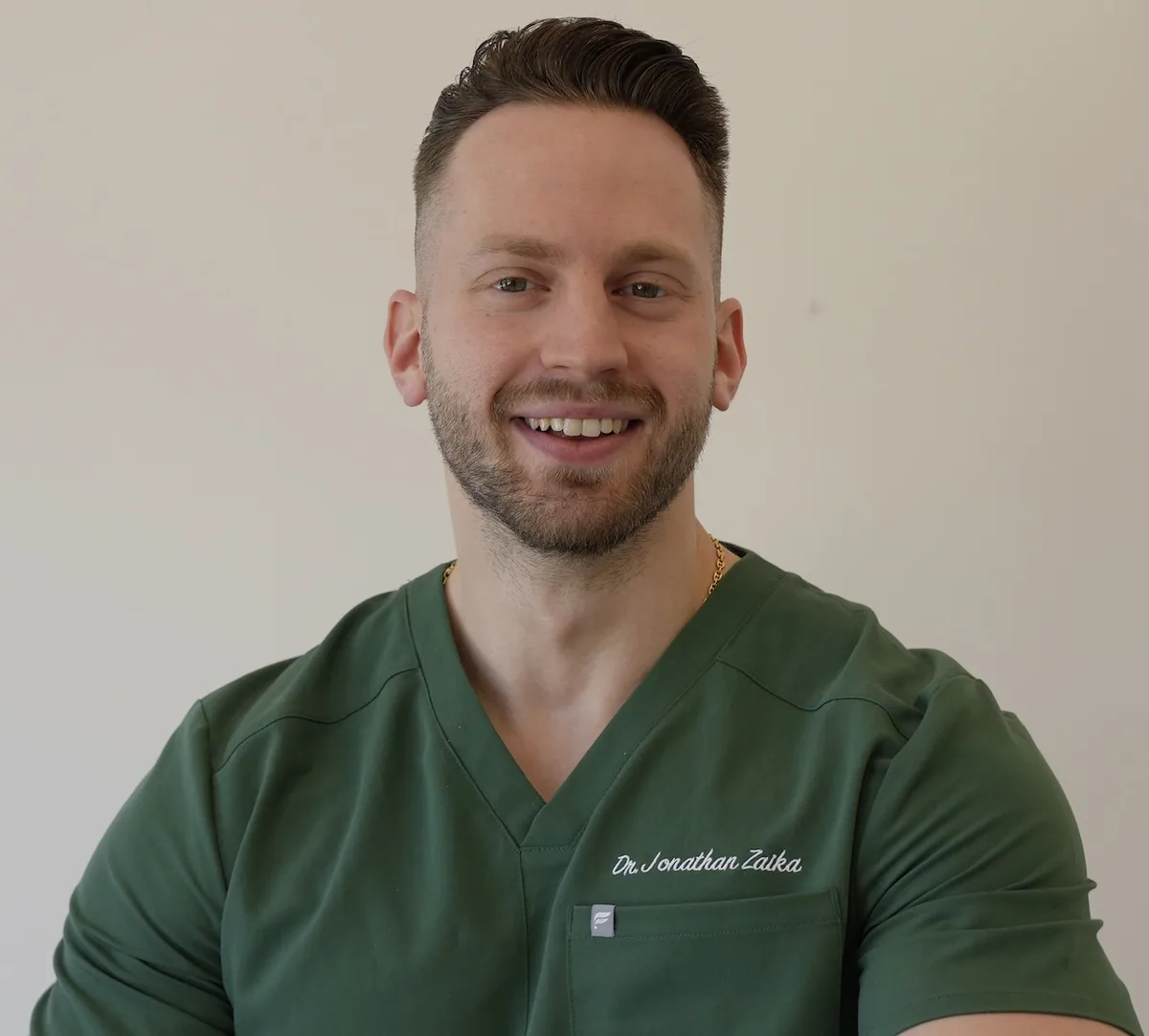
Education and training
New York University College of Dentistry, Doctor of Dental Surgery
Read more about Dr. Jonathan Zaika
Education and training
Stony Brook University School of Dental Medicine, Doctor of Dental Surgery (Summa Cum Laude); NYC Health + Hospitals/South Brooklyn Health, General Practice Residency
Read more about Dr. Stanley FaynblutRelated care
Visit us
Book online or call the office. A booking request is confirmed by our front desk; our team will contact you to confirm the time.
Get in touch
Have questions or need directions? We are here to help. Reach out or visit our office.
Call the office or send us an email and we will get back to you.